Tennis leg and osteopathy
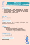
The tennis leg is characterized by the detachment of the medial gastrocnemius muscle (previously known as the medial gastrocnemius) from its common insertion with the soleus muscle on a membrane called the aponeurosis. In simple terms, the medial gastrocnemius and soleus are joined by a film-like membrane called the aponeurosis. During an episode of tennis leg, this aponeurosis ruptures, often resulting in an upward displacement of the medial gastrocnemius. It's not uncommon for a hematoma to form in the affected area.
A little anatomy
The calf is the large muscular part at the back of the leg between the knee and the heel. The calf is made up of 3 muscles:
- The medial gastrocnemius
- The lateral gastrocnemius
- The soleus
A little nomenclature
The term "tennis leg" is used because it frequently refers to a muscular injury in the leg of tennis players. However, this condition is not exclusive to this sport and is also found in other disciplines, particularly running.
It's important to distinguish tennis leg from tennis elbow, which is a tendinopathy affecting the elbow, not a muscular lesion of the leg.
Causes of tennis leg
The main causes of this pathology include:
-
Overload or overuse: Excessive use of the gastrocnemius muscle during sporting activities such as tennis, running, jumping, or other sports involving sudden, repetitive movements, can put excessive strain on the muscle, leading to injury.
-
Sudden, intense movements: Sudden movements such as rapid changes of direction, jumping or sprinting can cause muscle tears, especially if the muscle is not sufficiently warmed up or is already fatigued.
-
Aging and reduced flexibility: With age, muscles lose their elasticity and strength, making them more susceptible to injury, especially during intensive physical activity.
-
Lack of proper warm-up: Failure to warm up muscles properly before physical activity can increase the risk of injury, as cold, stiff muscles are more susceptible to injury.
-
Muscle imbalance: Weakness or imbalance between different leg muscle groups can increase the risk of calf injury.
-
Injury history: People with a history of calf or Achilles tendon injuries may be more susceptible to tennis leg.
Tennis leg often occurs unexpectedly, and can cause acute pain and swelling in the calf region. Preventing this condition involves proper training, a good warm-up and avoiding excessive overload on the gastrocnemius muscle.
Tennis leg symptoms
The tennis leg clinic, i.e. the clinical signs and symptoms associated with this injury, generally includes the following:
- Acute pain: One of the most common symptoms is a sudden, intense pain in the back of the leg, often described as a sensation of a blow or tear in the calf. This pain is typically felt during physical activity, particularly during movements that place heavy demands on the calf, such as running or jumping.
-
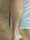
Edema andecchymosis: Swelling (edema) may occur in the affected area, often accompanied by bruising due to internal bleeding from ruptured muscle fibers.
-
Difficulty walking: Pain and swelling can make walking difficult and painful. The person may limp or be unable to stand on tiptoe on the affected side.
-
Deficit zone: On palpation, we can sometimes detect a deficit zone where the muscle has ruptured. This area may feel softer to the touch, due to the separation of muscle fibres.
-
Muscle weakness: There may be significant weakness in the calf, especially when attempting plantar flexion against resistance (such as pushing against an object with the foot).
-
Crepitus: In some cases, a crepitus sensation may be felt when palpating the area, due to air trapped in the tissue following rupture.
It's important to note that these symptoms may vary depending on the severity of the injury. In all cases, medical assessment is recommended to confirm the diagnosis and exclude other conditions, such as deep vein thrombosis or Achilles tendon rupture, which may present similar symptoms. Treatment and management of tennis leg will depend on the severity of the injury and the patient's specific needs.
Complementary examinations
Muscle ultrasound +++: Ultrasound is often the examination of choice for visualizing soft-tissue structures. It can detect muscle ruptures, hematomas, and abnormalities in the gastrocnemius muscle and Achilles tendon. Ultrasound is non-invasive and can provide detailed images of soft tissues.
Magnetic Resonance Imaging (MRI ): MRI can be used in more complex cases or to obtain a more detailed assessment of the injury. It is particularly useful for visualizing the internal structures of muscles, tendons and other soft tissues. MRI is more expensive and less accessible than ultrasound, but offers better image resolution.
Tennis leg: immediate treatment
Stop all sporting activity immediately: even if you feel able to continue, it's crucial to stop to avoid aggravating the injury. Rest your calf: It's advisable not to stress your calf. Avoid tensing it or walking normally, as this can be painful and damaging.
Apply ice for 12 minutes immediately after injury: There's no need to continue icing for the next few days, as this may interfere with the natural healing process and the inflammation that contributes to it. The absence of ice after the first few moments can speed up recovery.
Use a compression bandage : Apply light compression to the affected area for a few minutes, then maintain a moderately tight bandage to support the area without impeding circulation.
Avoid massaging the injured area for the first few days: Massaging can aggravate a potential hematoma and delay healing.
Elevate the leg: This can help reduce swelling and promote blood circulation in the injured area.
Tennis leg treatment
Osteopathy and tennis leg
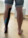
For a recent injury: The osteopath is qualified to manage injuries sustained during physical activity. He is competent to assess your injury and can apply strapping, sometimes in combination with taping, as shown in the image.
At a later stage: The osteopath's aim will be to treat any side-effects this injury may have had, not only on your calf, but also on the rest of your body. It is essential to wait at least 3 to 6 weeks to allow the injury to heal properly and ensure that the patient no longer limps or limps very little.
Other treatments: physiotherapy
Physiotherapy plays an essential role in the treatment and rehabilitation of tennis leg, a calf muscle injury. Here are the main steps and techniques used in physiotherapy for this condition:
-
Initial phase - Pain and inflammation management:
- Cryotherapy: Application of ice to reduce swelling and pain.
- Rest and elevation: Reduce activity to promote healing, and elevate the leg to reduce swelling.
- Compression: Use compression bandages to minimize swelling.
-
Recovery phase - Functional rehabilitation:
- Mobility exercises: Gentle movements to restore range of motion in the ankle and knee.
- Manual therapy: Gentle manipulation and mobilization to improve flexibility and muscle function.
- Muscle strengthening: Progressive exercises to strengthen the calf and surrounding muscles.
-
Advanced phase - Return to normal activity:
- Walking and running training: Gradually increase the intensity and duration of walking, then running.
- Proprioceptive exercises: Improve balance and coordination to prevent future injuries.
- Stretching: Stretching to improve flexibility and reduce muscle tension.
-
Recurrence prevention:
- Patient education: Advice on posture, lifestyle and warm-up techniques to prevent future injuries.
- Home exercise program: Specific exercises to maintain calf strength and flexibility.
-
Complementary Techniques:
- Electrotherapy: Use of electric current for pain management and muscle stimulation.
- Ultrasound: Used to promote tissue healing.
The physiotherapist adapts the treatment according to the severity of the injury, the patient's response to treatment, and his or her specific goals. Close communication between patient and therapist is essential for optimal recovery. It is important to follow the recommendations of the physiotherapist, and to avoid returning too soon to activities that could aggravate the injury.
Marie Messager
Osteopath D.O
2 rue Alexis de Tocqueville
78000 Versailles