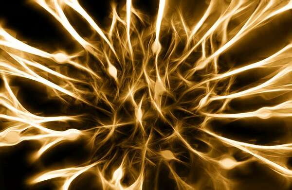
Presentation of the radial nerve
It is a mixed nerve, the most voluminous and it is mainly involved in the extension of the arm.
Origin of the radial nerve
The radial nerve (C6-T1) comes from the branch of divisor...
on 06/08/19
Presentation of the radial nerve
It is a mixed nerve, the most voluminous and it is mainly involved in the extension of the arm.
Origin of the radial nerve
The radial nerve (C6-T1) comes from the branch of divisor...
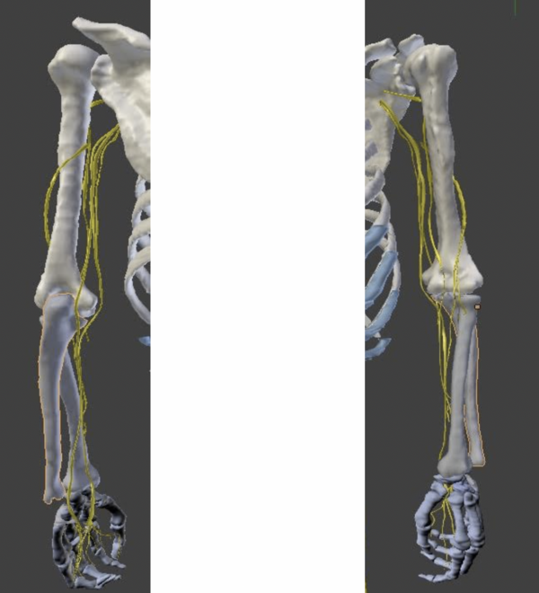
3D vector reconstruction of
Korean Visible Human
Left Upper Extremity Nerves
Title
3D vector modeling from anatomical sections of Korean Visible Human (KVH): recon...

3D vector reconstruction of
Korean Visible Human
Left Upper Extremity Nerves
References
DS Shin, MS Chung,JS Park et al. Portable Document Format File Showing the Surface Model...

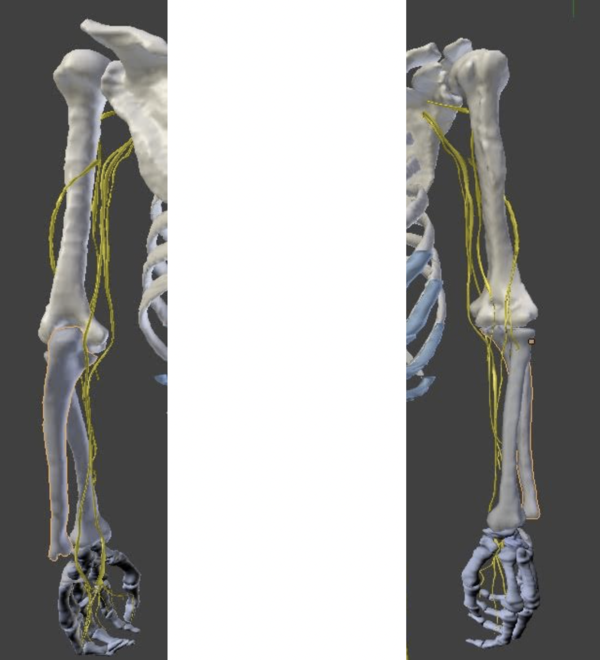
3D vector reconstruction of
Korean Visible Human
Left Upper Extremity Nerves
Conclusion
The Korean Visible Human (KVH) is a Korean project of 3D modeling of the human body in the...

3D vector reconstruction of
Korean Visible Human
Left Upper Extremity Nerves
Discussion
This research work on the nerves of the left upper limb and on the roots of the plexus of the...

3D vector reconstruction of
Korean Visible Human
Left Upper Extremity Nerves
Results
In total, the roots of the brachial plexus (from C5 to T1) as well as 6 nerves of the upper limb ...
3D vector reconstruction of
Korean Visible Human
Left Upper Extremity Nerves
Materials and methods
Selection of the body
The body chosen in our study was selected from all...

3D vector reconstruction of
Korean Visible Human
Left Upper Extremity Nerves
Table of Contents
1 Introduction and objectives
1.1 Objectives
1.2 State of the art
1.3 History

Presentation of the optic nerve II
The 2nd cranial nerve is the optic nerve. It is a sensory nerve that is responsible for vision.
40mm long, with a diameter of 4mm, the optic nerve is formed ...

Presentation and clinic of the Olfactory Nerve
The olfactory nerve or rather the olfactory nerves are sensory nerves.
They are responsible for conveying olfactory impulses. Thus, an attack on these nerves can cause...
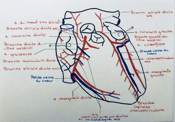
Arteries of the heart
The two coronary arteries of the heart, located under the epicardium, are the first branches of the ascending aorta.
Caliber: 3.5 mm.
The right coronary artery
Origin of the artery ...
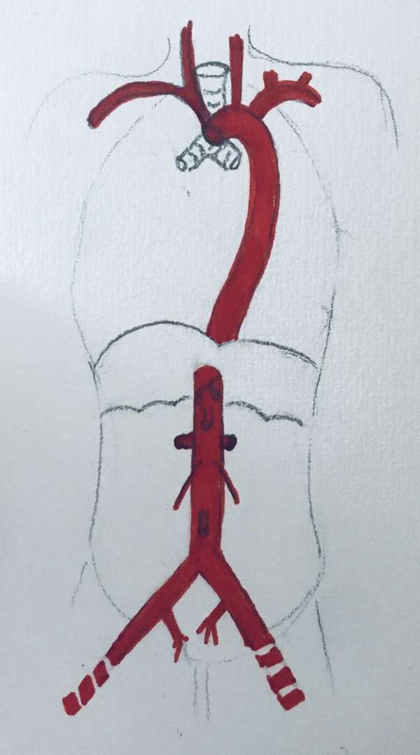
Presentation of the aorta
The aorta originates from the base of the left ventricle and ends at the level of the body of the L4 vertebra by dividing into 3 arteries:
left iliac artery right iliac artery a...
OSTEO VERSAILLES - Workshops
with Marie Messager
Osteopath Versailles
2 Alexis de Tocqueville Street
78000
Versailles
France
Building C3
Ground Floor
First door on the right
Viroflay, Le Chesnay, Vélizy-Villacoublay, Buc, Jouy-en-Josas, Vaucresson, Saint-Cyr-l'École, Chaville, Ville-d'Avray, Sèvres, La Celle-Saint-Cloud, Garches
Site design and SEO by Simplébo
![]()