Anatomy of the aorta
Presentation of the aorta
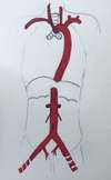
The aorta originates from the base of the left ventricle and ends at the level of the body of the L4 vertebra, dividing into 3 arteries:
- left iliac artery
- right iliac artery
- median sacral artery
Path of the aorta
The aorta runs obliquely up, forward and to the left for about 5 cm and thus forms the ascending aorta.
Then, it bends towards T4 and gives the aortic arch.
It descends against the left side of the spine to the 8th thoracic vertebra (T8) where it runs along the medial side of the spine to its termination. This is the descending aorta.
The descending aorta, crossing the diaphragm, changes from thoracic to abdominal aorta.
Dimensions of the aorta
The aorta presents a dilatation at its origin in front of the aortic valves which is called bulb of the aorta. Its caliber is 25 mm then about 19 mm. The latter decreases after the origin of the renal arteries to measure about 14 mm.
Structure of the aorta
The wall measures about 1.5 mm and has 3 layers or tunics (from the inside to the outside):
- Intima
- Media
- Adventine
The different parts of the aorta
1. The ascending aorta
Reports of the ascending aorta
Intrapericardial relationships:
- Forward: The right auricle
- Posteriorly: It is separated from the right pulmonary artery and the right atrium by the transverse sinus and the supra-arterial recess of the transverse sinus
- Right: The Lower Vault
Extrapericardial reports:
- Forward: The thymus
- Laterally: Mediastinal pleura and lungs
Collaterals of the ascending aorta
At its origin, the ascending aorta gives the right and left coronary arteries.
2. The aortic arch
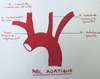
Presentation of the aortic arch
The aortic arch is almost horizontal and is located at the level of the 4th thoracic vertebra (T4).
Path of the aortic arch
The aortic arch runs obliquely backwards and to the left. It has 2 concavities; one directed downward and the other to the right.
Relationships of the aortic arch
Top side:
From front to back, we find:
- The brachiocephalic trunk
- The left common carotid artery
- The left subclavian artery
In front of the left brachiocephalic vein passes
Bottom side: it answers to :
- The bifurcation of the pulmonary trunk
- The left main bronchus
- The cardiac ganglia
Antero-left side: from front to back, we find
- The phrenic nerve
- The upper cervical cardiac branches
- The vagus nerve
- The left superior intercostal vein runs along this side behind the phrenic nerve
It is uncovered by the left mediastinal pleura
Posterior-right side: from front to back, it crosses
- The trachea
- The left upper tracheobronchial nodes
- The left recurrent laryngeal nerve
- The esophagus
- The thoracic duct
Collateral branches of the aortic arch
- Brachiocephalic trunk
- Thyroid artery ima (inconstant)
- Left common carotid artery
- Left subclavian artery
3. The thoracic aorta
Presentation of the thoracic aorta
The thoracic aorta is located in the posterior mediastinum and follows the aortic arch at the inferior border of T4. It continues with the abdominal aorta at the inferior border of T12 through the aortic hiatus of the diaphragm.
Path and relationships of the thoracic aorta
Forward:
- Left pulmonary root and ligament
- Left vagus nerve, top
- Aortico-esophageal pleural recess, below
Back:
- Thoracic spine
- Accessory hemi azygos vein
- Left great splanchnic nerve
Left: Left mediastinal pleura
Right:
- Lateral aspect of the thoracic spine
- Esophagus, top
- Thoracic duct
- Azygos vein, below
Collaterals of the thoracic aorta
- Right and left bronchial arteries
- Pericardial branches
- Esophageal branches
- Mediastinal branches
- The superior phrenic arteries
- Posterior intercostal arteries
4. The abdominal aorta
Presentation of the abdominal aorta
It is the terminal part of the descending aorta. It is located in the abdominal cavity.
Path of the abdominal aorta
It originates at the level of the aortic hiatus, opposite the lower border of the body of T12.
Located in the medial retroperitoneal space, it descends downward to the left of the midline to L4. It then terminates in 3 branches: the right and left common iliac arteries and the median sacral artery.
Reports of the abdominal aorta
In the aortic hiatus: The thoracic duct (posteriorly) and sometimes the medial root of the hemi-azygos vein.
In the abdomen: it is accompanied throughout its course by lumbar lymph nodes.
- Forward From top to bottom, we find
- The Omentary Scholarship
- The body of the pancreas
- The horizontal portion of the duodenum
- Hollow coves
- Back: Lumbar vertebrae L1 to L4
- On the right :
- Chlorine tank
- Thoracic duct
- Right celiac ganglion
- Azygos vein
- Inferior vena cava
- Right pillar of the diaphragm
- On the left :
- Left pillar of the diaphragm
- Left celiac ganglion
- Duodeno-jejunal angle
- Sympathetic trunk
Collaterals of the abdominal aorta
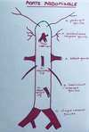
- The inferior phrenic artery
- The 5 lumbar arteries
- The celiac trunk: it is composed by
- The left gastric artery
- The common hepatic artery
- Splenic artery

- Superior mesenteric artery
- The middle adrenal artery
- Renal arteries
- Testicular and ovarian arteries
- The inferior mesenteric artery
Terminal branches of the abdominal aorta
- Common iliac arteries
- Medial sacral artery
Marie Messager
Osteopath in Versailles
78 - Yvelines