Hip abductor muscles
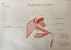
Abduction is a movement in which the thigh moves away from the axis of the body. When the fixed point is reversed, it is an elevation of the pelvis on the opposite side.
The movement is performed in the frontal plane and passes through the femoral head.
Theoretically, it is possible to abduct only one hip, but in practice abduction of one hip is automatically accompanied by equal abduction in the other hip and this is clearly seen from 30°.
The range of motion of abduction is between 35° and 45° and depends on age.
This movement is allowed by a certain number of muscles of the gluteal region and in particular by the middle gluteal muscle.
Superficial plane of the buttock area
The superficial plane includes the gluteus maximus and tensor fasciae lata (TFL) muscles.
Gluteus maximus muscle
Presentation
The gluteus maximus is the most voluminous muscle of the buttock
It is composed of a deep plane and a superficial plane and is abductor only by its highest fascicles.
Proximal Insertion
- Superficial plane: it is inserted by aponeurotic fibers in a common insertion on :
- on the outer side of the posterior ¼ of the outer lip of the iliac crest
- on the EIPS
- on the medial sacral crest
- deep plane: it is inserted by fleshy fibers in a common insertion on :
- the gluteal surface of the coxal bone behind the posterior gluteal line
- on the posterior aspect of the posterior sacroiliac ligaments
- on the lateral crest of the sacrum
- on the lateral edge of the sacrum and the coccox
- on the posterior surface of the sacro-tubal ligament
Path, belly
It is thick, diamond-shaped and its fibers are fasciculated (parallel), with an oblique path down and out.
Distal insertion
- Superficial plane: it ends with fascial fibers on the upper 1/3 of the posterior edge of the fascia lata
- deep plane: it inserts distally by fleshy fibers on the upper 1/3 of the lateral lip of the middle part of the acrid line and extends on the entire lateral lip of the trifurcation of the acrid line
Reports
- In front: gluteus medius muscle covered by the fascia lata and TFL (the superficial plane of the gluteus maximus, the fascia lata and the TFL form the "Faraboeuf gluteal deltoid".
- Back:
- synovial bursa
- the pelvi trochanteris
- the sciatic nerve
Action
- Static:
- contractile cushion provided with the presence of subcutaneous and deep synovial bursae.
- The amyotrophy of this cushion muscle facilitates the appearance of bedsores
- Dynamic: antigravidic muscle that deflects the thigh when walking:
- Thigh extension
- Lateral rotation
- Abduction from 45° F° onwards
Innervation: Inferior gluteal nerve (L5, S1, S2)
Fascia Lata tensor muscle
Presentation
The tensor fascia lata is an elongated muscle stretched from the hip bone to the tibia
Proximal Insertion
It is inserted thanks to :
- Fleshy fibers of the external face of the EIAS
- Aponeurotic fibers on the anterior 2/3 of the lateral border of the iliac crest
Path, belly
The fleshy fibers run downward and backward and then run into the anterior edge of the fascia lata and then become vertical at the level of the tibial iliac tract
Distal insertion
Normally, the termination of the TFL is on the fascia lata. The iliotibial tract ends :
- By a flattened tendon on the infra condylar tubercle (formerly gerdy's tubercle).
- Expansions to the femoral and leg fasciae
- Patellar expansion towards the sartorius forming the anterior knee cap
Reports
- The fleshy part is found between
- in front: the tendons of the femoral and sartorial rights
- backwards: the fascia lata
- on the surface : the skin
- in depth: the small buttocks
- The tendon is located in front of the femoral biceps. It is, externally, in contact with the lateral surface of the lateral condyle and the friction of these two structures can lead to a wiper syndrome
Action
- static :
- role of aponeurotic tensor
- at the thigh: with the gluteus maximus muscle, it is a proximal tensor of the thigh (does the work of a garter belt)
- at the knee: with the expansions of the biceps femoris muscles and the muscles of the goose paste, it is a proximal tensor of the leg (work of the support socks)
- Role of passive lateral support of the hip and knee during munipodal support
- Dynamic:
- At the hip: Flexion, Abduction, medial rotation
- At the knee: lateral rotation (it controls medial rotation) and locks the end of the extension
Innervation
Superior gluteal nerve (L4, L5, +/- S1)
In practice, it is when the 2 muscular parts of the gluteal deltoid work together in a balanced manner that the traction of the tendon is done in the longitudinal axis thus allowing the gluteal deltoid to perform a pure abduction.
Middle plane of the buttock area
The middle plane consists of the gluteus medius muscle.
Gluteus medius muscle
Presentation
Gluteal muscle stretched from the hip bone to the femur and triangular in appearance
Proximal Insertion
It is inserted by fleshy fibers
- on the midfield of the gluteal surface of the coxal bone between the anterior and posterior gluteal line
- the deep surface of the middle part of the gluteal fascia (fascia lata)
Path, belly
Fan-shaped, with lower top:
- the anterior fibers converge downward and backward
- the posterior fibers converge downward and forward
- it covers the small buttocks
- it runs along the upper edge of the piriformis muscle
Distal insertion
It ends with a tendon on the lateral side of the greater trochanter
Reports
- forward: the TFL
- backwards: the gluteus maximus (surface) and the piriformis (in depth)
- outside: the fascia lata
- inside : the small buttocks
Action
- static : Main role
- role of lateral support: active (during walking) of the hip during monopodal support. The failure of this function leads to Tredelenbourg's claudication. It is a hip limp with the pelvis tilting to the side opposite the affected gluteus medius.
- Dynamics: Hip abduction: The main abductor muscle is the gluteus medius: with its 40 cm cross-sectional area and 11 cm stroke, it has a power of 16 kgm (kilograms). It is very efficient because its direction is almost perpendicular to its lever arm.
Innervation
Superior gluteal nerve (L4, L5, S1)
Deep plane of the buttock area
It includes the
- the small gluteal muscle (gluteus maximus)
- the pelvi trochanteric muscles, from top to bottom:
- piriform
- upper twin
- internal shutter
- lower twin
- external obturator: non-abductor of the thigh
- femoral square: not abductor of the thigh except at 110° of flexion
Small buttock muscle
Presentation
Gluteal muscle stretched from the hip bone to the femur and triangular in appearance
Proximal Insertion
It is inserted by fleshy fibers on the anterior field of the gluteal surface of the coxal bone in front of the anterior gluetal line
Path, belly
Fan-shaped, with lower apex:
- the front fibers are vertical
- the posterior fibers become horizontal
It runs along the upper edge of the piriformis muscle and then covers the coxofemoral joint
Distal insertion
It ends with
- a tendon on the anterior aspect of the greater trochanter
- an expansion on the superior iliofemoral ligament
Relationships
It is located between the TFL on the surface and the coxofemoral joint in depth. It is on the same plane as the piriformis, which is located behind it.
On this same plane, the gluteus minimus, piriformis, obturator internus, twins, quadratus femoris and adductor magnus muscles form what is known as the "Sioux Cuff".
Action
- Static: stabilization of the coxofemoral joint
- Dynamic:
- Hip flexion, abduction: it is three times less powerful on the abduction than the gluteus medius
- Rotation by its anterior fibers
Innervation
Superior gluteal nerve (L4, L5, S1)
Piriform
Presentation
Pelvitrochanteric muscle stretched from the sacrum to the femur, triangular in appearance
Proximal Insertion
It is inserted by fleshy fibers on the anterior surface around the2nd and3rd sacral foramina and on the adjacent lateral part
Path, belly
Triangular, it goes down, forward and outward
- Its course is initially endopelvic in contact with the anterior sacral roots
- Then it exits the pelvis through the large sciatic incision
- It is finally extra pelvic covered by the gluteus maximus
Distal insertion
It ends with a tendon on the superior aspect of the greater trochanter
Reports
At the endopelvic level, it is related to
- forward with the roots of the sciatic nerve
- below with the pudendal plexus
It divides the great sciatic incision into 2 spaces: supra and infra piriformis.
At the extra pelvic level, the pyramidal is located under the gluteus maximus, above the obturator internus and the twins.
Action
- Static:
- stabilization of the coxo femoral
- coaptation of the sacroiliac joint: it moves the sacral base down and out
- Dynamic:
- Lateral hip rotation up to 45° of flexion
- At 45° hip flexion, it is an abductor
- Cybernetic role (automatic balancer): return spring which brings in intermediate position (anteversion/retroversion).
Innervation
Piriformis nerve (S2)
Internal obturator and the upper and lower twins
Presentation
Pelvic trochanter muscles, stretched from the hip bone to the femur and triangular in appearance
Proximal Insertion
It is inserted by fleshy fibers on :
- the circumference of the obturated foramen on the internal surface of the coxal bone
- the upper (for the upper twin) and lower (for the lower twin) edges of the small sciatic incisure
Pathway, abdomen
The 3 muscles form a "pelvic triceps" with an intra and then extra pelvic course.
Wide at the beginning, the fibers converge backwards and outwards towards the small sciatic incisure.
The fibers exit the pelvis through the small sciatic incision, making a 90° reflection.
They are separated from the bony edge by a bilobed synovial bursa which ensures a non-traumatic sliding.
They then move forward and outward
Distal Insertion
Their distal insertions end in a common tendon on the medial aspect of the greater trochanter anterior to the trochanteric fossa
Reports
At the intra pelvic level, the internal obturator is between
- outside: the Inner Obturator Membrane
- inwards: the levator ani
- top: the vasculo-nervous bundle
- below: the pudendal canal
At the level of the small sciatic incisure, the pelvic triceps is located in an osteo-fibrous canal limited by :
- above: the sacrospinous ligament and the sciatic spine
- in front: the bony edge
- below: the ischial tuberosity
- backwards: the sacro tuberal ligament
At the extra pelvic level, it is located in the Sioux cuff between the piriformis at the top and the femoral square at the bottom
The sciatic nerve passes backwards
Action
- Static: stabilization of the coxofemoral joint
- Dynamic:
- Cybernetic role (automatic balancer): they participate in the gemello-obturator hammock that supports the pelvis in relation to the femoral heads
- Lateral hip rotation up to 90° flexion
- At 90° of flexion, they are hip abductors
Innervation
- Nerve of the internal obturator and the superior gastrocnemius (L5,S1 , S2) for the internal obturator and the superior gastrocnemius
- Nerve of the lower twin and the femoral square (L4, L5, S1) for the lower twin

Successive engagement of the abductors
Depending on the degree of flexion, in unilateral support, the pelvis will be stabilized by different muscles.
- By the TFL when the hip is in extension
- When the pelvis is a little less tilted backwards, the center of gravity falls behind the hips and the gluteus maximus begins to kick in.
- The gluteus medius in neutral position, i.e. when the pelvis is balanced in the anteroposterior plane.
- From the moment when the pelvis tilts forward, (thus in flexion), we see successively intervene :
- The large buttocks
- The 45° piriformis
- The internal obturator and the 90° twins
Marie Messager,
Osteopath in Versailles Chantiers
Yvelines - 78